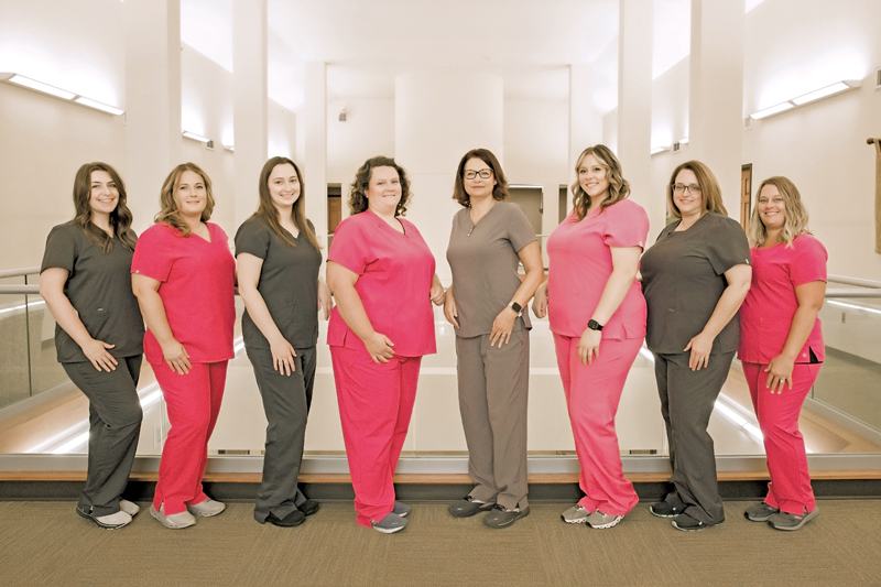
Our radiologists and technologists provide comprehensive radiology procedures. These procedures are performed by or under the supervision of certified radiologists in a team atmosphere where leading-edge technology is the standard. Certified radiologists provide the interpretations for all the comprehensive radiology procedures. Our in-house radiologist along with our certified, trained professional technologists provide exceptional service to our patients.
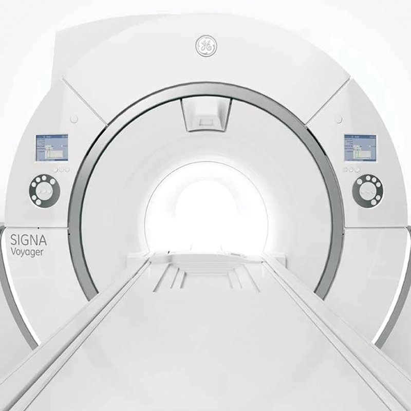
Magnetic Resonance Imaging (MRI)
MRI service is offered daily.
Appointments available: Monday - Friday, 7:00 am - 4:00 pm.
MRI is a way of allowing your physician to see inside your body without surgery or radiation. MRI uses a strong magnetic field and radio waves. There are no known biological hazards associated with MRI. The MRI magnet has an opening through which the table moves; this ensures that the necessary part of the body will be scanned.
STH offers Wide Bore MRI
- Largest bore opening on the market for patient comfort.
- Exam room includes customizable lightingand sound during your exam.
- State-of-the-art digital imaging and table weight limit of 500 pounds.
Nuclear Medicine
Nuclear Medicine service is offered daily. Appointments are available Monday - Friday, 7:00 am - 2:00 pm.
Nuclear Medicine studies document organ function and structure in contrast to conventional radiology, which creates images based on anatomy. Many nuclear medicine studies can measure the degree of function present in an organ, oftentimes eliminating the need for surgery. Some of the most common procedures are myocardial perfusion, which aids in the diagnosis of Coronary Artery Disease (CAD), or blockages in the arteries that supply blood to your heart muscle. Other common studies include lung scans for the detection of blood clots; bone scans that can detect fractures, tumors, infections, etc.; and Gallbladder scans to detect obstructions of chronic infection.
Ultrasound
Ultrasound service is available Monday - Friday by appointment.
STH also features state-of-the-art ultrasound equipment. More than just a way to see a fetus in the womb, the ultrasound machine can be used for many functions. Some of those are looking at gallbladders, clots in leg veins, breast lumps and checking neck arteries following strokes. It is also used for guiding biopsies and performing echocardiograms (studies of the heart).
3D Mammography
Available Monday - Friday by appointment.
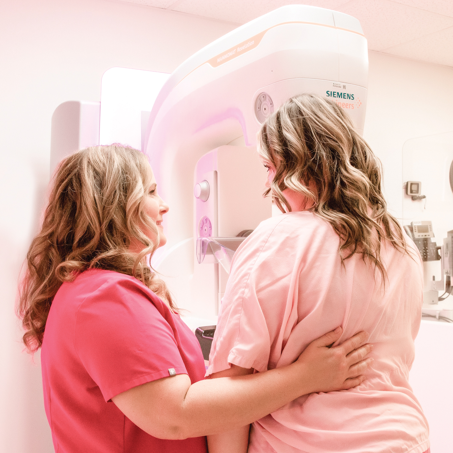
The 3D mammography machine allows for high image quality mammography giving every woman her best chance for an early and accurate diagnosis.
The technology features Flex compression plates with self-adjusting tilt depending on the thickness and shape of the breast to provide a more comfortable and uniform compression over the breast surface. Mammography is one of the most federally controlled modalities in Radiology. All of the mammography technologists require advanced certifications.
Dexa Scan
Available Monday - Friday by appointment.
DEXA is the most superior technology for measuring bone health. A bone density test measures the health of bones. Osteoporosis is a potentially crippling disease that affects over 75 million people worldwide. Osteoporosis is one of the most preventable diseases today. The lifetime risk for developing osteoporosis is 1 in 2 Caucasian women.
Intervention Radiology
Available Wednesday and Friday by appointment.
A sub-specialty of radiology that involves the treatment of numerous disease processes with minimally invasive, image-guided techniques. These procedures include arthrography, biopsies, drainages, therapeutic joint injections, ultrasound-guided breast needle localization, and Sentinel Lymph Node Localizations.
Computed Tomography (CT)
Available Monday - Friday by appointment.
Computed tomography shows organs of interest at selected levels of the body. CT scans are the visual equivalent of bloodless slices of anatomy, with each scan being a single slice. CT examinations produce detailed organ studies by stacking individual image slices. The scans are produced as the x-ray beam circles around the patient.
Computed Tomography or CT scan is also known as a CAT (computerized axial tomography) scan. This is a unique examination because it combines the use of X-rays and a computer to produce clear, sharp pictures within your body. The images of your body are divided into slices, much like slices from a loaf of bread. The radiologist is able to see bones and soft tissues within your body using a CT scan. This diagnostic tool provides a quicker, more accurate diagnosis of many medical conditions that are very difficult to detect with regular X-rays.
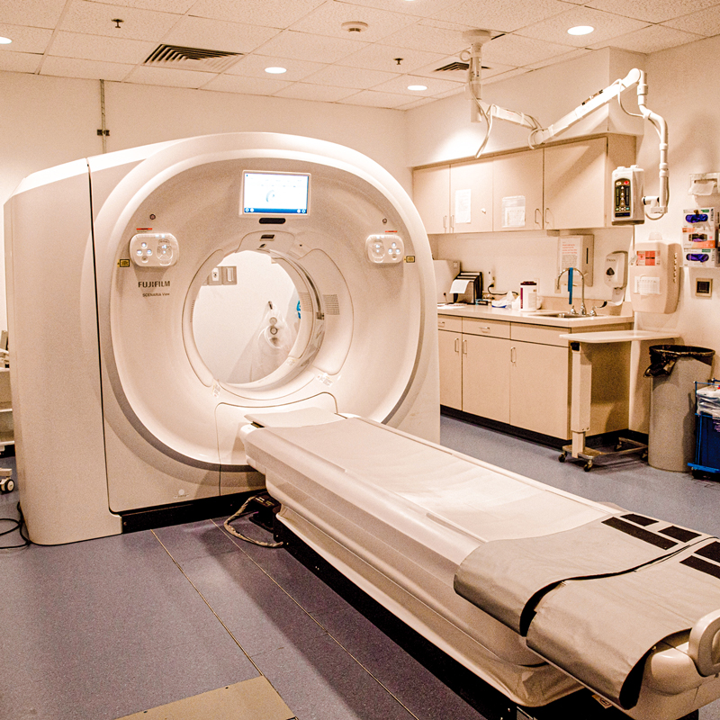
SCENARIA View CT
- More spacious 80cm opening is larger than similar CT systems to reduce anxiety
- Lateral Shift Table allows you to remain centered and secure
- The table accommodates patients up to 550lbs
- The patient table lowers to 20” from the floor to allow easy access
- Advanced technology allows fast scanning for minimal scan time
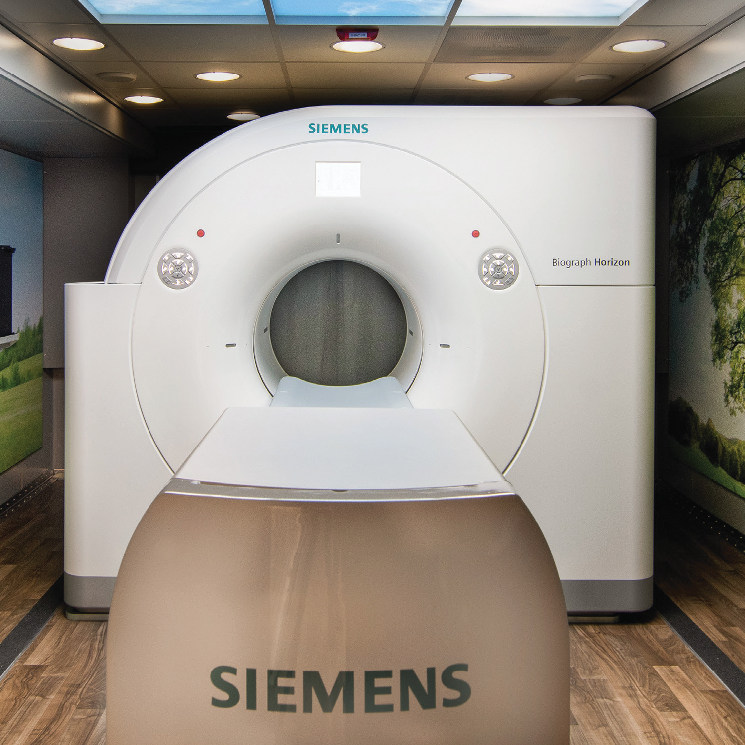
PET/CT (Positron Emission Tomography/Computed Tomography Imaging)
For Scheduling Call: 618-740-1644
PET/CT combines the functional information from a Positron Emission Tomography (PET) exam with anatomical information from a Computed Tomography (CT) exam in one single exam. A PET scan detects changes in cellular function–specifically, how your cells are utilizing nutrients like sugar and oxygen. Since these functional changes take place before physical changes occur, PET can provide information that enables your physician to make an early diagnosis. The advantage of CT is its ability to take cross sectional images of your body. These are combined with the information from the PET scan to provide more anatomic details of the metabolic changes in your body. The PET exam pinpoints metabolic activity in cells and the CT exam provides an anatomical reference. When these two scans are fused together, your physician can view metabolic changes in the proper anatomical context of your body.
A PET/CT study not only helps your physician diagnose a problem, but it also helps your physician predict the likely outcome of various therapeutic alternatives, pinpoint the best approach to treatment, and monitor your progress.
